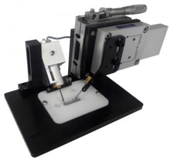
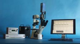
Intact papillary muscle isolated from hearts, located halfway between single cell and whole heart experimental research, offers special benefits.
FEATURES
- In muscle preparations, simultaneously measure calcium and force
- Capable of handling a range of sample types (papillary muscle, trabeculae, EDL, soleus, etc)
- Stiffness can be described using a precise length motor
- Stretch protocols that may be programmed
The intact papillary muscle can be utilized to study cardiac function in a multicellular context with an intact 3-dimensional myofilament lattice, in contrast to single isolated myocytes, which constitute the smallest fully functional model system. Furthermore, unlike whole heart investigations, papillary muscle's contractile properties can be assessed without relying on external factors like vascular tone. Moreover, papillary muscle enables measures that are too challenging or impossible to carry out in complete hearts. Similar to how skinned preparations cannot simultaneously detect force production, quantify tissue force, measure intracellular calcium dynamics, or perform a stretch-release technique, intact muscle preparations can.
These measurements are made possible by the IonOptix Intact Muscle Chamber. A strong force transducer and programmable length controller can be easily mounted to intact ventricular papillary muscle. While the specific force transducer permits electrical excitation straight through the prepared muscle, chamber fluid flow enables for temperature regulation and continual oxygenation of tissue. The muscle ends are knotted with simple platinum "Omega" clips, which slide onto platinum hooks. Because the platinum is inert and permits electrical conductance, there is no electrolysis or disturbance of biology.
IonOptix Intact Muscle Chamber has been tested and used with a variety of skeletal preparations, including EDL, soleus, and engineered tissue, in addition to cardiac tissue.
IonWizard, which enables control of experiment acquisition parameters and data processing, can be used to detect the fluorescence of calcium-sensitive dyes like Fluo-4, Fura-2, and/or Indo-1 when used in conjunction with an Intact Muscle Chamber System and an IonOptix Calcium and Contractility System. It is also simple to analyze entire skeletal muscles, such as the soleus of the mouse.
SPECIFICATIONS
- Range of sensitivity: 100 mN (typical, more sensitive force transducers available with 5 mN detection limits)
- > 500 Hz is the system resonant frequency.
- Depending on the application, there are two types of length controllers: voice coil (5mm travel, 20 nm resolution, slower reaction) or a piezo motor (50 µm travel, 0.10 nm resolution, faster reaction)
COMPONENTS
To provide a complete set of simultaneous fluorescence and contractility (i.e., force development) measurements, Intact Muscle Chamber Systems can be integrated with standard Calcium and Contractility components. Systems could come with a programmable length controller or a sensitive force transducer, depending on your particular application.
Microscope
Inverted microscopes with IonOptix configurations offer the perfect platform for measurements combining photometry and dimensioning. In order for these microscopes to function as the imaging component within IonOptix Calcium and Contractility Systems, they have been designed to contain numerous proprietary IonOptix optical components and connectors. Depending on your application, pick between the Olympus IX73 or the Motic AE31.
Light Source
Your unique research requirements guide the selection of the fluorescence excitation light source, which can be set up for practically any dye or dye combination. Monochromatic LEDs, the extremely quick HyperSwitch, or the more reasonably priced MuStep are all possibilities.
Sensors
The Intact Muscle Chamber system's sensors are in charge of converting cellular signal into data. A photomultiplier tube and the high-speed MyoCam-S3 digital camera are used to assess whole-cell fluorescence photometry and cell contractility, respectively. The Cell Framing Adaptor enables you to frame a cell of interest and decrease background fluorescence for cleaner fluorescence signals.
Force Transducer
Typically measuring muscular force growth, the reliable force transducer has a sensitivity range of 0-100 mN.
Stimulator
In order to give tissue acute stimulation during excitation-contraction experiments, the field stimulator is a crucial component. For complex sequencing and arrhythmic stimulation, use the MyoPacer EP instead of the MyoPacer for straightforward stimulation procedures.
Tissue Chamber
Muscle strips can be mounted on inert platinum hooks with ease, and inlet and outlet perfusion flow are both made possible by the specially built muscle chamber. It works with the majority of inverted microscope stages.
Interfaces
The FSI connects the hardware and software in the Calcium and Contractility system, enabling simultaneous control of calcium and/or contractility measurements as well as inputs and outputs for external hardware.
Length Controller
Based on length range, resolution, and frequency response, programmable length control can be customized to fit your studies.
SOFTWARE
The core IonWizard program, which displays force data and length controller position in trace recordings, is responsible for the majority of the muscular system acquisition. You might also include the software modules PMTAcq for fluorescence recording and the Advanced Signal Generator for precise, protocol-based muscle length control.


