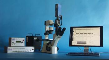
Myocyte Calcium and Contractility Systems
The system measures calcium levels and contractility in isolated myocytes. It is a unique development that allows to combine fluorescence photometry with digital measurement of cell geometry.
- Overview
- Links
A complete bench-top solution for measuring digital cell geometry and acquiring fluorescence photometry simultaneously to investigate the function of myocytes
IonOptix is pleased to provide systems that are at the forefront of the business for monitoring myocyte calcium and contractility. All systems enable simultaneous acquisition of digital cell geometry measurements and fluorescence photometry, which is unique.
System parts have been created and built to function as a whole within an integrated workstation. Acquiring data this way is precise and synchronous. The systems are completely integrated and set up on-site, allowing the researcher to get started right away after delivery. IonOptix' systems give researchers the ability to obtain cell length and sarcomere spacing along with their calcium measurements, which is more than a general fluorescence system can do.
Your preparations are used when we install complete systems to help you get going as soon as possible. Additionally, IonOptix provides limitless phone and email help for the duration of your system should you require it.
FEATURES
- Complete, turn-key system
- Measurements of cell shrinkage and calcium photometry that are simultaneous and coordinated
- Suitable for a range of cell types, including iPSC-CMs, skeletal muscle, and adult cardiomyocytes
- Rapid, precise calcium
- Intuitive transient analysis is included in the software package
- Adaptable to your unique study requirements
COMPONENTS
Depending on your application, systems are available with any of our three stimulation light sources: LEDs, the HyperSwitch, or the µStep. With acquisition speeds up to 1000Hz, single, dual excitation, and dual emission photometry are all available to cover all potential fluorescence probes.
The light source and probe have an impact on the photometry acquisition speeds. Our MyoCam-S3 offers measurements of cell dimensions at up to 1000 Hz, as well as simultaneous capture of cell and sarcomere length. Additionally, up to 4 channels of 12-bit analog data can be acquired concurrently using IonOptix' Fluorescence System Interface at a preset rate of 1000Hz.
IonOptix can connect the system to an already installed microscope or include a microscope with the system. The manufacturer also offers a variety of auxiliary equipment, such as heaters, chambers, and digital field stimulators, to finish the physiological measurement system.
Microscope
Inverted microscopes with IonOptix configurations offer the perfect base for measurements combining photometry and dimensioning. In order for these microscopes to function as the imaging component within Calcium and Contractility Systems, they have been designed to integrate various unique IonOptix optical components and connectors. Depending on your purpose, pick either the Olympus IX73 or the Motic AE31.
Light Source
Your unique study requirements guide the selection of the fluorescence excitation light source, which can be set up for almost any dye or dye combination. Monochromatic LEDs, the extremely quick HyperSwitch, or the more reasonably priced MuStep are all possibilities.
Sensors
Cellular signals are converted into data by the sensors present in the Calcium and Contractility system. A photomultiplier tube and the high-speed MyoCam-S3 digital camera are used to assess whole-cell fluorescence photometry and cell contractility, respectively (PMT). The Cell Framing Adaptor enables you to frame a cell of interest and decrease background fluorescence for better fluorescence signals.
Cell Stimulator
To give cells a quick stimulation, the field stimulator is a crucial part of excitation-contraction investigations. For complex sequencing and arrhythmic stimulation, use the MyoPacer EP instead of the MyoPacer for straightforward stimulation procedures.
Cell and Tissue Chambers
During experiments, cells must be kept in a suitable chamber environment. During your investigations, you can stimulate and perfuse cells using the FHD Chamber and the C-Stim Chamber, and you can precisely adjust the temperature of the chamber bath with the Cell Microcontrols MTCII.
Interfaces
The FSI connects the hardware and software in the Calcium and Contractility system, enabling simultaneous control of calcium and/or contractility measurements as well as inputs and outputs for external hardware.
SOFTWARE
IonWizard combines the display of analog voltages, cell length statistics, and fluorescence traces and photos into a user-friendly interface. It uses ratio, ion, or linear calibration calculations to assess raw data.
Calculations for fluorescence use a wide range of background-subtraction methods. Trace averaging, composite pictures, and transient analysis are examples of specialized functions that eliminate typical unnecessary effort.
Integrating with other programs is made simpler by the clipboarding and exporting of trace and image data. Using context-sensitive online support and user-specific templates frequently boosts productivity.
You can also visit site of the manufacturer.


