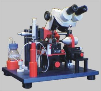
SI-H Standard Muscle Research
Integrated system for mechanical and optical registration of intact or dissected muscle tissue. Supports many modern methods of muscle physiology research.
- Overview
A study device called SI-H Standard Muscle Research combines mechanical and optical recording of whole muscles and dissected muscle fibers.
A number of contemporary research techniques in the area of muscle physiology are incorporated into a single instrument by the SI-H Standard Muscle Research system. The system of linear motors, laser diodes, photometers, perfusion cuvettes, and thermostats can be added to the KG mechanosensors on a solid platform. Complex experiments benefit from this flexibility.
SI-H Standard Muscle Research is capable of conducting the following five crucial muscular physiology experiments:
- Skeletal muscle responses to electrical stimulation or tetanus are characterized by measuring the mechanical characteristics of the contracting and relaxing muscle fibers.
- Jerk force measurement, kinetic analysis, time to maximum force, semi-"viscosity," relaxation's start curve, and diastolic force change.
- In order to assess the mechanical properties of the muscle fibers, a linear motor must be connected to a control unit. These properties include sagging, isotonic relaxation, "viscosity" of relaxation, vibration tests, post-load contractions, and eccentric contractions.
- By observing diffraction from a laser diode, it is feasible to measure sarcomere length simultaneously.
- A gradient-creation function is added to allow for the automated analysis of force-pCa ratios and the impact of calcium on individual muscle fibers.
Features:
- Collecting and analyzing data
- Design flexibility and modularity
- A pair of cuvette tables
- Construction with stainless steel (stainless steel, anodised aluminium, plastic)
- The optical register has a lifetime warranty
Accessories
- Linear motor for the study of changes in muscle length
- Ergometer for postload contraction in cardiac muscle and eccentric contraction
- Stimulator and thetanizer for stimulation of intact muscles
- Photometer for the study of intracellular calcium.
- Cuvette system for the study of contraction during rapid cooling
- Rapid temperature change system for individual muscle fibers
- Software controlled gradient creation for calcium studies
- Laser diode for the diffraction study of sarcomeres in muscle and muscle fibres
- Laser controlled perfusion system for accurate measurement of weak forces
- Optional accessories like load cells, amplifiers, data acquisition systems (ADC)


