- Our e-mail:
RFLSI III Laser Speckle Imaging System
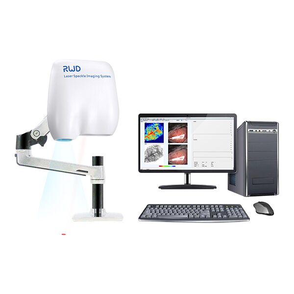
This product is discontinued.The RWD RFLSI III laser speckle imaging system allows visualization of blood perfusion and has advanced software to help monitor and record blood perfusion in real time.
- Overview
- Specifications
- Links
The RFLSI III is a reliable blood perfusion imaging system. In conjunction with cutting-edge software, it aids researchers in real-time monitoring and recording blood perfusion of any exposed tissues or organs for microcirculation investigation, allowing them to instantly examine the quantified data, cut down on experiment time, and rapidly receive results.
The LSCI (Laser Speckle Contrast Imaging) technique is the foundation of RFLSI III. For the purpose of studying microcirculation, it can be used to watch and record blood perfusion of any exposed tissues or organs thanks to its benefits of non-contact, fast frame rate, and high spatial resolution.
This device is intended for pre-clinical microcirculation studies on topics such as mesentery, lower limbs, and ischemic stroke. Blood perfusion photos and videos (4.2 million pixels), as well as quantitative data for perfusion unit and vessel diameter, are included in the multi-output.
Applications:
- Cerebral blood perfusion monitoring
- MCAO model assessment
- Cortical spreading depression observation
- Hind-limb ischemia research
- Skin burn/skin flap transplantation
- Organ microcirculation observation
Advantages
Accurate Data Measurement
With accuracy up to 0.1 PU and simple controls, stable laser output allows for dependable and consistent measurement over the course of minutes, hours, and days.
HD Picture & Video in Full-Frame
The field of microcirculation imaging is made possible by high-definition cameras with full-frame 2048x2048 resolution, which allow you to observe the blood vessel terminals.
A Rapid Frame Rate
You may capture quick changes every second and capture more specific blood vessel alterations following treatment with a high-speed camera (up to 120 FPS).
Data for Quantitative Visualization
Monitoring the entire field and quantitative analysis. Vascular perfusion volume, vessel diameter, vessel angle, and other outputs are included in the multi-output, and offline state analysis is also possible.
Ensured to Run without Issue
USB 3.0 is supported. Data gathering and the production of a film showing blood perfusion happen instantly.
Simple Operation
No need for a specialized dark environment thanks to unique non-contact technology. Quickly obtain perfusion data and video using the autofocus function.
You can also visit site of the manufacturer.

We are a distributor of RWD Life Science Inc. in Estonia, Latvia, Lithuania and Ukraine, Kazahstan and Europe