- Our e-mail:
Optopatcher
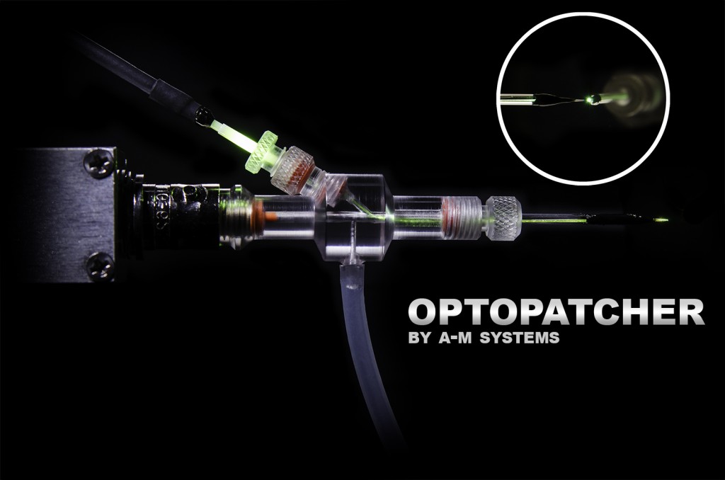
Micropipette holder with integrated fiber optic, allowing the combination of optogenetic stimulation and electrophysiological registration from the cell.
- Overview
- Links
Are you interested in enhancing your patch clamping methods with the power of optogenetics?
When using an in-vivo patch-clamp procedure, the Optopatcher® offers unsurpassed accuracy in providing optical stimulation. An optical fiber and an electrode are both housed in the holder, allowing for simultaneous patch-clamp recording and optogenetic activation.
This is a considerable benefit for non-slice techniques including whole-cell patch-clamping, spike/single-unit recording, in vivo recording from intact animals, and LFP recording.
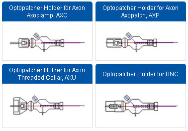
The Optopatcher® can be ordered with the most popular patch clamp connectors, including Heka and A-M Systems' basic BNC connectors as well as Axon's Threaded Collar, Axopatch, and Axoclamp connectors. Capillary glasses with a diameter of between 1.2 mm and 2.0 mm can be supplied to fit it. Please get in touch with us for bespoke diameters.
The Optopatcher comes in two different models. Users can attach their light source to the short internal optic fiber of the Optopatcher's STANDARD configuration using a 1.25mm ceramic sleeve. The Optopatcher's CONTINUOUS setup employs a single continuous fiber to connect directly to your light source instead of an internal optic fiber.
Each Standard Optopatcher® Includes:
- One starter fiber
- Replacement seals
- One mating sleeve for the ferrule
The Standard Optopatcher is user-friendly since the fiber optic cable may be unplugged, allowing for easy removal from the rig for patch pipette replacement. By removing the junction between the internal Optopatcher fiber and the fiber from the light source, the Continuous Optopatcher offers 10 times higher light transmission to the target tissue.
Each Continuous Optopatcher® Includes:
- Replacement seals
The optical fiber must be properly trimmed to its final length for any form of Optopatcher® in order for it to fit within the patch pipette. The fiber end did not require any specific tip treatment, according to earlier research by the Optopatcher® developers, for efficient light transmission. If the fiber end is placed close to the pipette's end and is blunt cut and unpolished, it may cause biological reactions. If your lab doesn't already have one, we advise first-time Optopatcher® buyers to additionally buy one of our ruby blade knives.
You can also visit site of the manufacturer.

We are a distributor of A-M Systems, LLC in Ukraine, Kazahstan and Europe