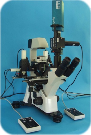
MyoStretcher System
MyoStretcher is the most sensitive system on the market, specifically designed to measure the contraction force of isolated intact cardiomyocytes.
- Overview
- Links
Record Force from Isolated Cardiac Myocytes!
The next-generation MyoStretcher System, which now uses ground-breaking New Optical Force Transducer, the OptiForce, has been released, and IonOptix is happy to announce it. It is now the most sensitive technology available for measuring forces from isolated intact myocytes! It was created exclusively for force measurement in cardiomyocytes!
The MyoStretcher was created with an emphasis on reliability, simplicity, and convenience of use. The MyoStretcher comes with everything needed to stretch isolated myocytes and measure force. Along with the sensitive force transducer, motorized micro-manipulators, and all component fittings, IonOptix also provides an optional piezo-electric translator for programmable stretching, a kit to make it easier to attach glass rods to the MyoStretcher arms, and an alternative set of patented concave glass rods designed to enhance the attachment of isolated myocytes.
Use the MyoStretcher to investigate:
- Accurate diastolic calcium
- Auxotonic and isometric contractions
- Length-dependent activation
- Force???velocity relationship
- Workloops in single cells
The MyoStretcher is a convenient modular add-on for your existing IonOptix Myocyte Calcium and Contractility System or it can be built as a stand-alone system. As always, every system includes on-site setup and instruction so you can start using your new tools as soon as possible.
FEATURES
- Measure calcium and shortening while being under physiologic strain
- Down to sub-nN resolution, measure the force development in a single myocyte
- Programmable stretch protocols
- Work loops can replicate the heart cycle thanks to a fast-feedback length motor
COMPONENTS
Depending on the particular research application, MyoStretcher systems can be highly customized. For passive stretch studies, only MyoTak and micromanipulators with glass rods are required. Add the piezoelectric motor for programmable length control and the incredibly sensitive OptiForce transducer for force readings. There is a stable post configuration available to minimize signal noise for extremely sensitive applications, such as Work Loops. Additionally, the MyoStretcher is a modular add-on for Calcium and Contractility devices that allows for simultaneous measurement of force, calcium release, and cell shortening.
Microscope
Inverted microscopes with IonOptix configurations offer the perfect base for measurements combining photometry and dimensioning. In order for these microscopes to function as the imaging component within Calcium and Contractility Systems, they have been designed to integrate various unique IonOptix optical components and connectors. Depending on your purpose, pick either the Olympus IX73 or the Motic AE31.
Light Source
Your unique research requirements guide the selection of the fluorescence excitation light source, which can be set up for practically any dye or dye combination. Monochromatic LEDs, the extremely quick HyperSwitch, or the more reasonably priced MuStep are all possibilities.
Sensors
Cellular signals are converted into data by the instruments present in the Calcium and Contractility system. A photomultiplier tube and the high-speed MyoCam-S3 digital camera are used to quantify whole-cell fluorescence photometry and cell contractility, respectively. The Cell Framing Adapter enables you to frame a cell of interest and minimize background fluorescence for cleaner fluorescence signals.
Cell Stimulator
To give cells a quick stimulation, the field stimulator is a crucial part of excitation-contraction research. For advanced sequencing and arrhythmic stimulation, use the MyoPacer EP instead of the MyoPacer for straightforward stimulation methods.
Interfaces
The FSI connects the hardware and software in the Calcium and Contractility system, enabling synchronous management of calcium and/or contractility measurements as well as inputs and outputs for external hardware.
Cell Chambers
During studies, cells must be kept in a suitable chamber environment. It is simpler to orient cells for mechanical loading with the rotational version of the FHD chamber that is offered for MyoStretcher systems.
Rail Configuration
An affordable rail alternative that works with the majority of microscope types is available for straightforward MyoStretcher configurations.
Stable Post Configuration
The stable post configuration offers a very safe platform for your MyoStretcher measurements in studies where stability is crucial, like work loops.
OptiForce
The IonOptix OptiForce is a ground-breaking optical fiber interferometry-based force transducer created as a key component of our MyoStretecher system and specifically created to identify the nanoscopic forces from single isolated cardiac cells. With detection limits as low as nanoNewtons and a frequency response above 3kHz, it offers two key advantages over current technologies: sensitivity and speed.
Length Controller
Piezo motor
With the piezoelectric motor for length control, you can set wave to wave amplitude and duration changes to create a unique stretch waveform and define cell length changes as percentage or absolute changes.
SOFTWARE
Users of the MyoStretcher can combine all necessary components into one user-friendly interface by using IonWizard. Users can activate the Advanced Signal Generator to stretch cells in accordance with any desired waveform, measured in absolute cell length or percent change, in addition to the straightforward contractility options.
You can use MultiClamp to simulate the cardiac cycle by clamping at a specific pre-load and after-load level, and IonWizard will be fully equipped to handle high-content characterization of myocyte physiology.
The MyoStretcher System is linked to five components in addition to IonWizard.
You can also visit site of the manufacturer.


