- Our e-mail:
Movable Objective Microscope?® Sutter MOM?®
The Movable Objective Microscope is a versatile optical microscope that combines the functions of a light-field, fluorescence microscope and a confocal two-photon microscope.
- Overview
- Specifications
- References
- Links
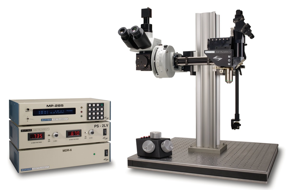
The Movable Objective Microscope® (MOM®) is a two- or three-photon microscope that, when used in conjunction with the right laser, can image deeply inside living specimens. The Sutter MOM became the first scope to offer rotation and movement in three dimensions, enabling the specimen to stay flat and stationary. The Sutter MOM is utilized by many renowned imaging laboratories all around the world, and Sutter team continuously collaborates with clients to modify the design to meet their evolving requirements.
MOM Opto-mechanical Design
The MOM is made up of two separate microscopes. An Olympus vertical illuminator, Sutter Xenon arc lamp, and camera mount make up the wide-field portion of the microscope, which provides conventional epifluorescence. The optical channel for moving the excitation laser light from the table up into the scanning galvanometric mirrors, expanding the beam through the scan lens, and directing into the rear of the objective is provided by the two-photon side of the microscope. After two-photon excitation, a dichroic mirror just above the objective directs the released photons into the detecting route. The main body of the microscope can be easily accessed before imaging since it travels backward on a rail system.
The objective spins around the X axis and moves in the X, Y, and Z directions. Regardless of position or orientation, the microscope can maintain efficient excitation light delivery to the rear aperture of the objective thanks to two movable mirrors. You may be sure that the X, Y, and Z movements are smooth, precise in scale, drift-free, and highly reproducible because they are the same as those in Sutter MP-285 micromanipulator. These movements make it possible to record Z-stacks and mosaic images of extensive tissue regions without the use of a moving stage.
It is possible to rotate the objective away from its usual vertical position thanks to the horizontal light path. This rotation enables the MOM to be quickly transformed from an upright to an inverted microscope, with the objective being positioned at any angle between 0 and 180 degrees. It is possible to image non-horizontal surfaces and volumes thanks to this positional freedom.
MOM Scanning Systems
Scanning methods for multiphoton microscopes have seen numerous changes over the past ten years. When large aperture, high NA objectives became accessible, larger aperture scanners became necessary. The use of a resonance scanner facilitated quicker imaging. Both resonant-galvo and resonant galvo-galvo systems are now available in two-photon scopes. New technology may be bolted onto older, existing scopes with little modification thanks to the Sutter MOM, which was created concurrently with these modifications. Numerous 3 mm galvo scanner original scopes have been replaced with 6 mm galvo scanners or resonant/galvo scanners. For instance, any new MOM system can include the Vidrio RMR scanner (a resonant galvo-galvo scanning system) or current MOM scopes can have it retrofitted.
Imaging Software
Sutter started selling the MOM Computer System and Software in 2011. (MCS). Prior to the creation of this program, the majority of users relied on ScanImage or MPScope to produce scanned images. Although customers appreciated that the MOM would work with open source freewares, there appeared to be a market for a commercial package as well. MCS continues to provide a straightforward, user-friendly package at a cost comparable to that of commercial and open-source software. The most recent version of MScan, 3.0, is compatible with Windows 10. The large (1-2 hour) data files that can be recorded in the MCS proprietary data file structure are used in a recent paper. (Consider Kuhn, 2020)
ScanImage freeware, the two-photon imaging program created by Karel Svoboda and associates, has always worked with the MOM®. The tremendous commitment from the ScanImage community is one of the factors enabling the MOM platform to exist in its current form. In 2014, Vidrio took over as the main platform for ScanImage support and new development. Customers who want the most recent features and premium service can get Vidrio ScanImage Premium from Sutter with pleasure. ScanImage Basic is a starter system that comes with a year of service. The free ScanImage software is still accessible, but it does not offer assistance. To connect the MOM and other scanning microscopes to ScanImage Premium, ScanImage Basic, or the freeware version, Sutter offers packages that include the essential data gathering hardware. In addition, Sutter offers Vidrio's hardware, such as the vDAQ acquisition system, RMR scanner, and computers that are ScanImage ready.
The following items are all included in Sutter MOM packages, with the exception of the laser and objective:
- Suitable tube and scan lenses for two- or three-photon imaging
- XY galvonometric (3 mm or 6 mm) or resonant scanners from Cambridge Technology (resonant-galvo or resonant galvo-galvo systems both with 5 mm mirrors)
- R6357 multialkali or H10770PA-40 (GaAsP) products are examples of Hamamatsu photomultiplier tubes (PMTs). (Other PMTs are available; Sutter is a certified Hamamatsu reseller.)
- Power supplies for PMTs: Sutter PS-2 (dual channel high-voltage power supply for conventional PMTs) or Sutter PS-2/LV (dual channel low-voltage power supply for H10770PA-40 or other PMTs with built in high voltage). Power supplies can be ordered with remote turn on/shut off for PMT gating
- Hamamatsu, Sigmann, or FEMTO pre-amplifiers
- Data acquisition: National Instruments PXI FPGA, Vidrio vDAQ, or National Instruments PC based Multifunction I/O
- Conoptics Pockels Cells for laser intensity control
APPLICATIONS
- In vivo two-photon imaging
- In vivo three-photon imaging
- Electrophysiological recording and imaging (culture, large in vivo preparations, etc.)
- Non-horizontal surface microscopy
- Simultaneous retinal stimulation and two-photon microscopy1
- Whole animal imaging
- Immunology
- Embryology
FEATURES
- Objective travels 22mm in X, Y, and Z.
- For imaging of volumes and surfaces that are not horizontal, the objective rotates around the optical axis.
- Adaptable open platform architecture
- Resonant or conventional XY scanners from Cambridge Technology
- Detector system with two or four channels, Hamamatsu PMTs, and preamplifiers
- Dual channel Sutter PS-2/PS-2LV PMT power supply
- Scan and tube lenses that are compatible with two or three photons
- *New* Light Block keeps ambient light, photostimuli, and visual stimuli out of the detector's field of view.
- Data gathering systems from National Instruments and Vidrio Technologies
Travel
- 22 mm on all three axes
Resolution
MP-285 controller
- Low: 0.2 µm/step
- High: 0.04 µm/step
MPC-200 controller
- 0.0625 µm/step
Maximum Speed
MP-285 controller
- 2.9 mm/sec
MPC-200 controller
- 5.0 mm/sec
Long Term Stability
- 1-2 µ/hour
Drive Mechanism
- Precision worm gear capstan drive
Communication
- MP-285: RS-232 Serial
- MPC-200: USB
Electrical
- 115/230 volts
- 50/60 Hertz power line
LAMBDA LS 300W XENON ARC LAMP
Lamp Life
- 1,000 hours (500 hour warranty)
- Longer life depends on application
Electrical
- 115/230 volts
- 50/60 Hertz power line
PS-2/PS-2LV PMT POWER SUPPLY
Electrical
- 115/230 volts
MDR-3/MDR-6/MDR-R SCAN DRIVE CONTROLLER
Electrical
- 115/230 volts
- 50/60 Hertz power line
![]()

The instrument was used in these investigations:
- Eyecup scope-optical recordings of light stimulus-evoked flourescence signals in the retina
Euler et al
Pflugers Arch, 2008
You can also visit site of the manufacturer.

We are a distributor of Sutter Instrument in Estonia, Latvia, Lithuania and Ukraine, Kazahstan and Europe