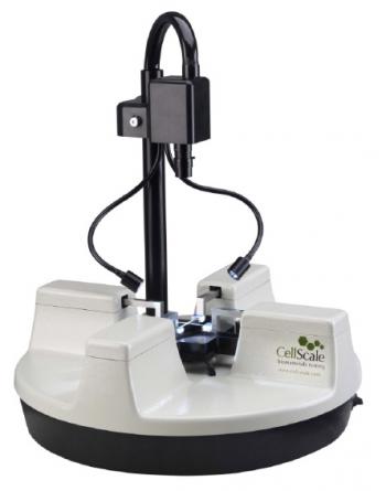
BioTester ??У Biaxial Tester
Biotester allows you to monitor the contraction or stretching of tissue on two axes, recording a video picture synchronized with the work of sensors. Specialized software allows you to build histograms of contractions and observe the movement of individua
- Overview
- Specifications
Fully equipped biaxial test systems built specifically for biomaterials
Given that biomaterials have directionally aligned microstructures, biaxial testing is essential for comprehending their mechanical characteristics. Biaxial testing has been made simple by the BioTester systems so that users can concentrate on outcomes rather than testing.
Key Features
- 4 actuators with high performance and inline load cells
- For quick and reliable specimen mounting, there are many attachment options available, including the unique BioRakes.
- High resolution imaging and methods for measuring strain from images
- Integrated media bath with temperature control
- Real-time feedback user interface program with full functionality for simple, cyclic, relaxation, and multi-modal testing
BioTester 5000
- The standard system comes with a full feature set
- Long stroke enabling grip separation of up to 80mm
BioTester 3000
- Numerous alternative features and a modular design
- More accessibility for imaging and other instruments due to small size
Specimens & Mounting
BioRake Sample Mounting System
Each tine is electrochemically sharpened so that it may easily pierce tissue samples of any hardness or fragility. To enable simultaneous piercing of all 20 connection sites, each set is firmly fastened to a shared base. The BioRakes are magnetically mounted to make it simple to switch between the BioRake, Balanced Pulley, and Clamp mounting methods as well as to remove them for cleaning or replacement.
Samples are placed and brought into position for testing using the manual lift mechanism, and pressure is then applied to embed the hooks in the tissue. Hence, the sample mounts quickly and is prepared for analysis. The mounting is reliable, precise, and simple.
To accept specimens ranging in size from 3 to 15mm, BioRakes are available with tine spacing options from 0.7mm to 2.2mm.
Balanced Pulley Mounting System
On each side of the specimen, there are two double-ended bespoke suture hooks utilized to make four attachment points. Each suture is maintained at the same tension during the test thanks to a two-stage stainless steel pulley system.
The pulley mechanisms are magnetically mounted to make it simple to switch between the BioRake, Balanced Pulley, and Clamp mounting methods as well as to remove them for cleaning.
Clamp Mounting System
The attachment sites, which are by nature weaker than the base material, can be removed from the specimen's gauge region by using a cruciform specimen. The specimen can be loaded and held securely using the clamps.
In order to make switching between the BioRake, Balanced Pulley, and Clamp mounting methods quick and simple, the stainless steel clamping mechanisms are installed over the same brackets used for the other attachment systems.
Moreover, unique clamp designs can be created to match your tissue's needs in terms of clamp force and clamping surface.
Software
The BioTester's software module for setup, operation, and data gathering makes it easy to carry out pre-established or specially designed test protocols.
For easy access and adjustment, test parameters including displacement magnitudes, durations, and data/image collection rates are defined in a tabular format. Each axis can have its own independent displacement and force regulated testing specifications. Once a protocol has been defined, a template system is utilized to quickly reload the required test parameters.
During the test setup, the software interface gives the user access to live imagery as well as current force, position, and temperature data. In order to enable user monitoring of the test progress, the program offers real-time results graphing and a live video feed while the test is in progress.
Everyone who has used this equipment will be happy to tell you how user-friendly and practical the program is.
BioRake Sample Mounting System
Biaxial testing involves a lot of analysis and interpretation of data.
The BioTester has a software module for image analysis that may be used to study and examine test photos and provide insightful quantitative and qualitative data. For presentations, the same module can be used to export.avi films or photos with data overlays.
Throughout the test, the BioTester's high definition camera provides a clear view of the specimen. The software module automatically time-correlates the collected photos (at a user-specified frequency) with the force displacement data in order to provide the user with instant access to all test results.
The software module's image tracking capability makes it simple and quick to analyze test photos in order to identify in-plane motions of one or more locations on the specimen surface. Instead of depending on strains calculated from grip motion, this tracking information offers a direct measurement of specimen strain. It is feasible to study the strains in various locations of the object by following a grid of points.


